Community Posts
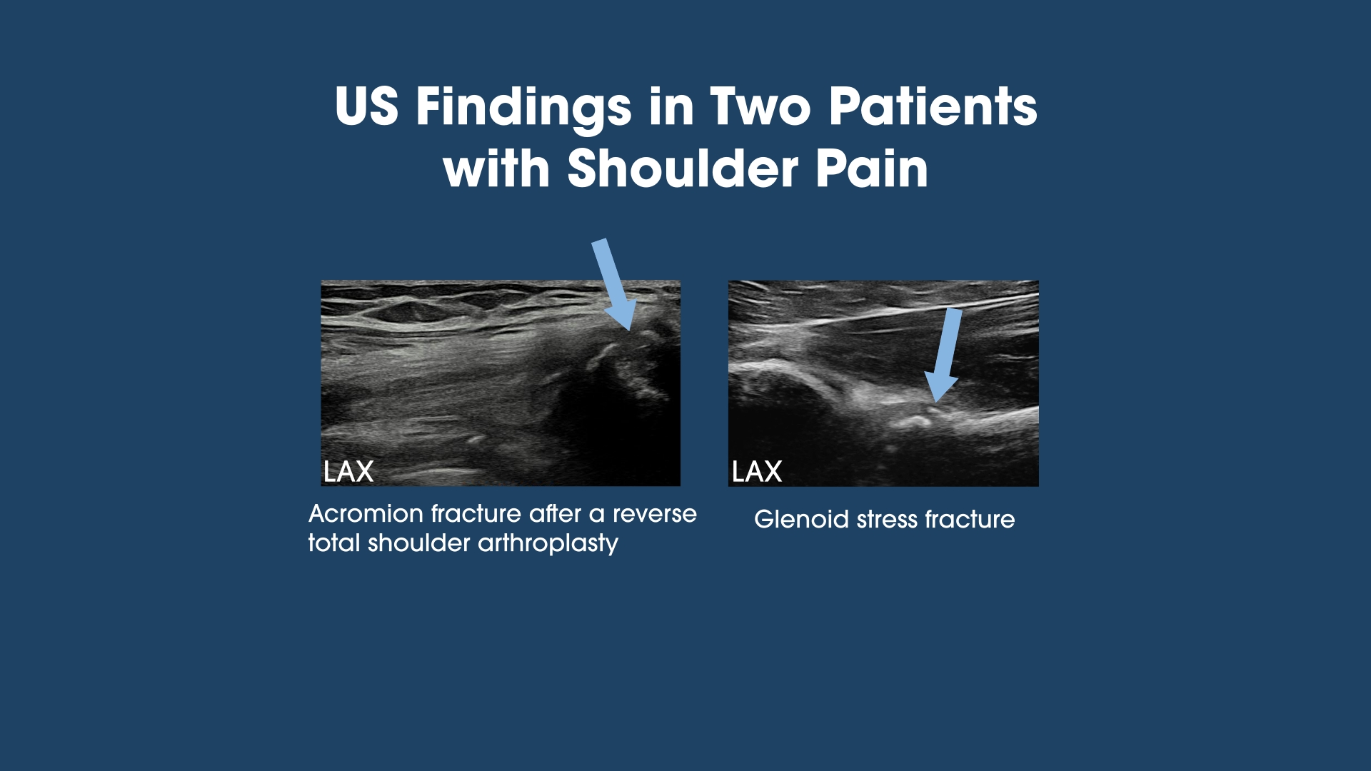
Ultrasound Detection of Two Unexpected Shoulder Fractures READ MORE »

Welcome to UltimateMSK
Community Posts
Ultrasound Detection of Two Unexpected Shoulder Fractures READ MORE »
Community Posts
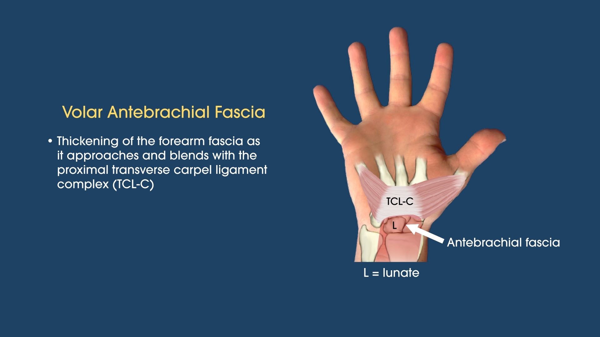
The Antebrachial Fascia in the Carpal Tunnel Evaluation READ MORE »
News
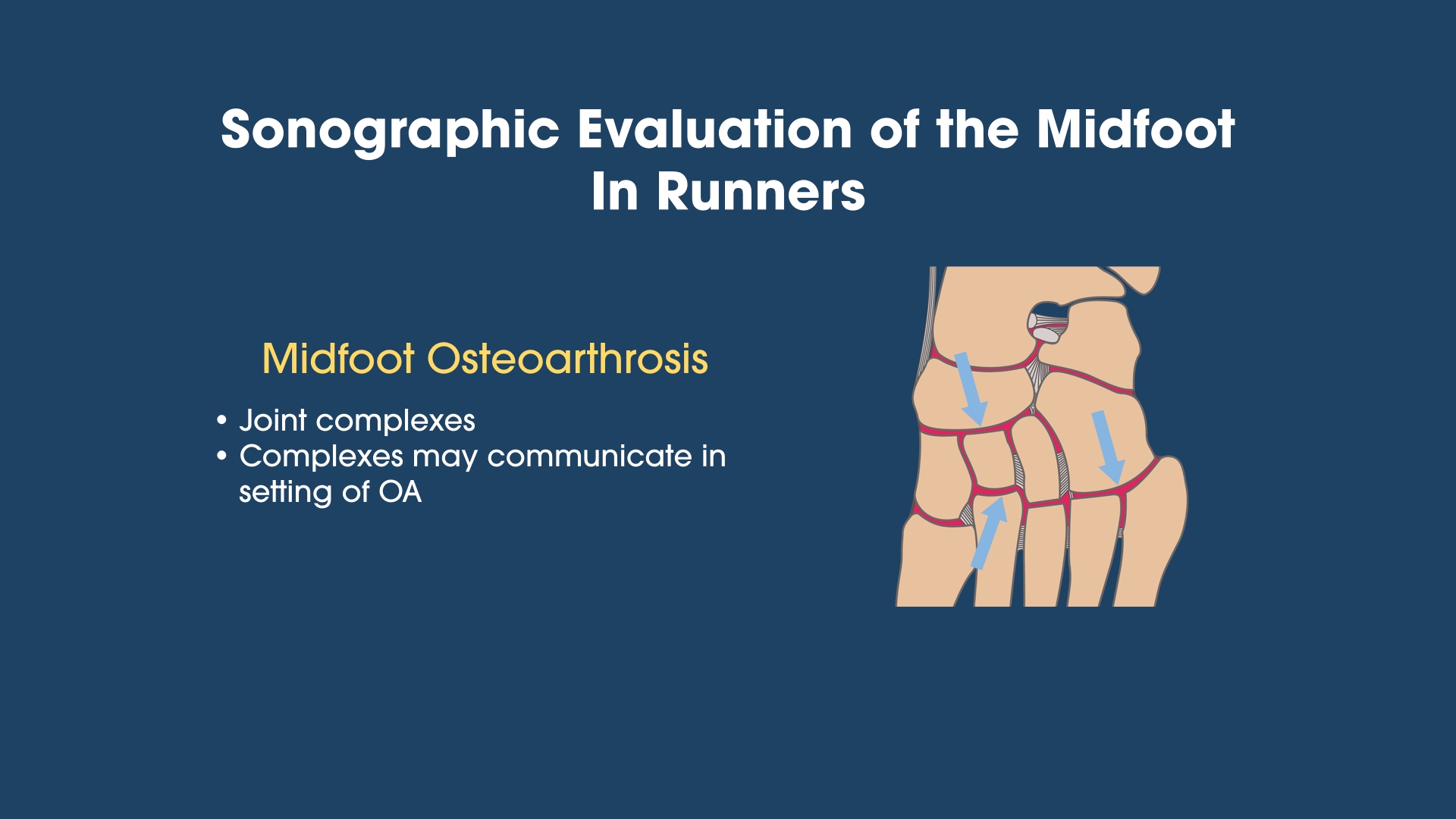
Sonographic Evaluation of Foot and Ankle Injuries in Runners READ MORE »
News
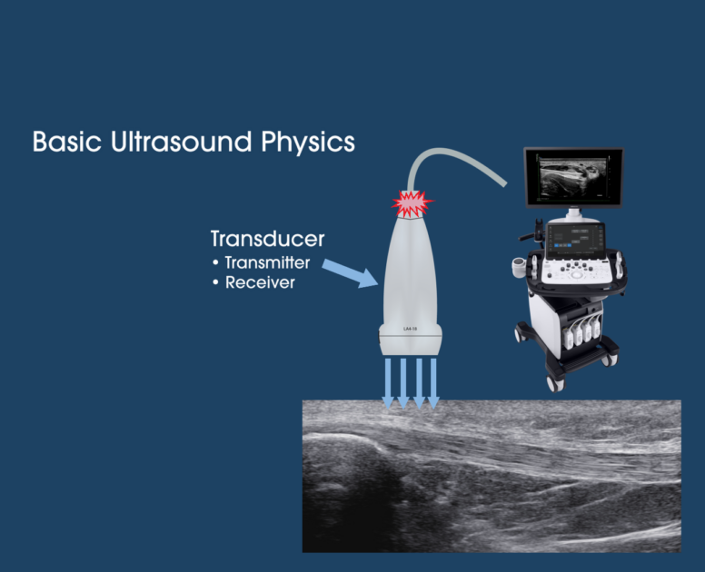
Ultrasound Basics READ MORE »

News
Introductory Video READ MORE »
Discover the ultimate destination for MSK ultrasound training with UltimateMSK, a cutting-edge, web-based platform designed to elevate your learning experience. Offering a comprehensive suite of teaching modules, UltimateMSK empowers you to master musculoskeletal ultrasound at your own pace through its flexible, subscription-based format.
Each module includes free CE credits, ensuring you gain valuable certifications while advancing your skills. Beyond training, UltimateMSK fosters a vibrant community where subscribers and visitors alike can connect, collaborate, and grow. Share images, discuss cases, and ask questions in a supportive space dedicated to musculoskeletal ultrasound enthusiasts. Join UltimateMSK today and experience a dynamic, learner-focused environment that combines expert instruction with unparalleled community engagement.
UltimateMSK is your essential resource for advancing your MSK US education. Whether you’re just starting out or already integrating MSK US into your practice, UltimateMSK provides the tools and insights to help you reach your goals.

Step By Step Approach
Each teaching module will present the scanning protocol, review the pertinent anatomy and demonstrate a live scan, and discuss the spectrum of pathology.
Board Certified Instructor
Doug Hoffman, M.D., RMSK, is the founder and director of the MSK Ultrasound Department for a large multi-specialty group. He has also held national leadership roles in MSK US education.
CE Credit Availability
Each module includes CE Credits available to a wide range of health care providers at no extra costs.
Available Pathways

Comprehensive Diagnostic
The Comprehensive Diagnostic Pathway is designed for all levels of US knowledge and skills. The modules are broken down into those of the upper extremity and lower extremity.
Podiatric
The Podiatric Pathway focuses on the foot and ankle and will be an invaluable resource to those just beginning their MSK US journey to the advanced practitioners.


Orthopedics & Sports Medicine
Dr. Hoffman practiced sports medicine and orthopedics for nearly 20 years before integrating MSK US into his practice 15 years ago.
Testimonials
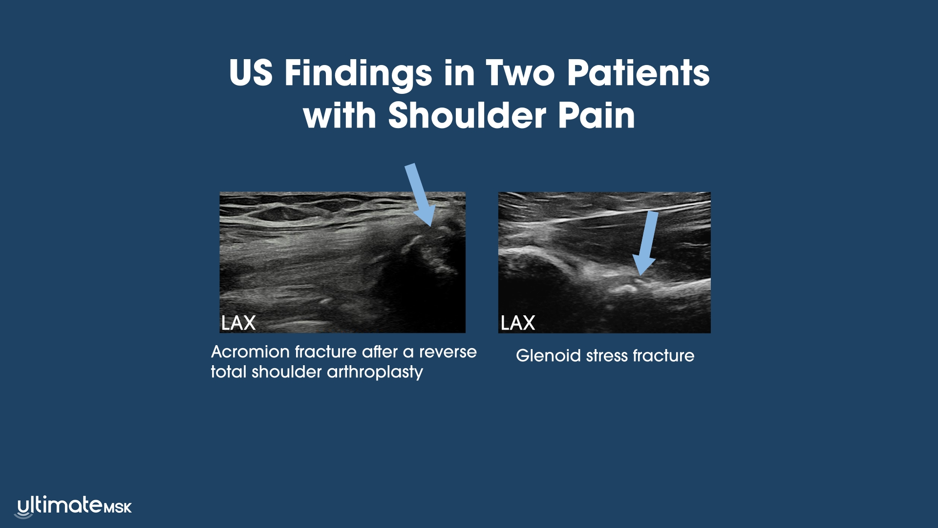
Ultrasound Detection of Two Unexpected Shoulder Fractures
The Antebrachial Fascia in the Carpal Tunnel Evaluation

Plantar Hindfoot Pain after a Hilly Trail Hike
UltimateMSK is a comprehensive web-based platform organized into multiple teaching modules. The subscription-based format allows you to study at your own pace, ensuring that you can practice and revisit the material as often as you need to achieve proficiency in musculoskeletal US. Each module includes free CE credits. In addition to the learning modules, UltimateMSK offers a dedicated space for collaboration. Visitors to the site, whether they have a subscription or not, can ask questions, share interesting images or cases, and engage with a growing community of musculoskeletal US enthusiasts. UltimateMSK’s goal is to foster a collaborative learning environment that enhances the experience for all of us.
The mission of UltimateMSK is to promote musculoskeletal ultrasound (MSK US) education and support the growth of the MSK US community through both didactics and collaboration. Proficiency in MSK US is a continuum that requires both individual learning and community engagement. UltimateMSK strives to become a leader in MSK US education by offering comprehensive and easy-to-understand learning modules, ongoing case examples, interesting images, and sharing the experiences of others in the field.
UltimateMSK is also developing advanced learning modules on topics such as the brachial and cervical plexus, advanced shoulder US including the pectoralis major and latissimus dorsi muscle-tendon units, the deep gluteal space, and a rheumatology module for non-rheumatologists. Additionally, UltimateMSK will foster ongoing collaboration with MSK US practitioners across all disciplines and levels through case discussions, Q&A sessions, webinars, and various other postings related to MSK US.
Orthopedics & Sports Medicine
Dr. Hoffman has over 30 years of experience in sports medicine and orthopedics. He is the founder and director of a dedicated MSK Ultrasound Department at a large multispecialty medical system and is actively involved with MSK US education and research.
Dedicated MSK US clinical practice
Peer-reviewed publications
MSK US education leadership positions
Testimonials
Douglas Hoffman, MD
Dr. Hoffman has over 30 years of experience in sports medicine and orthopedics. In 2012, after completing a mini-fellowship in Europe with leading MSK radiologists, he founded and has since directed the MSK Ultrasound Department at a large multispecialty healthcare system, fostering collaboration between the orthopedic and radiology departments. His practice is exclusively focused on MSK ultrasound, encompassing diagnostic imaging, ultrasound-guided injections, and advanced ultrasound-guided procedures. Dr. Hoffman is also actively involved in MSK ultrasound education and research.
Please check out the sample videos from the completed Hamstring, Forefoot, and Plantar Hindfoot Modules, as well as the nearly completed Wrist Module.
Overview
Anterior Ankle into the Midfoot
Lateral Ankle
Medial Ankle
Posterior Ankle – The Achilles Tendon
Overview
Anterior Elbow
Lateral Elbow
Medial Elbow
Posterior Elbow
Overview
Forefoot Introduction
Forefoot General Approach Sample
Forefoot Protocol Sample #1
Forefoot Protocol Sample #2
Anatomy and Live Scan
Forefoot Anatomy and Live Scan – Dorsal Sample #1
Forefoot Anatomy and Live Scan – Dorsal Sample #2
Forefoot Anatomy and Live Scan – Dorsal Sample #3
Forefoot Anatomy and Live Scan – Plantar Sample #1
Forefoot Anatomy and Live Scan – Plantar Sample #2
Forefoot Anatomy and Live Scan – Plantar Sample #3
Pathology
Forefoot Pathology Part 1 – Joints and Metatarsal Stress Fractures Sample #1
Forefoot Pathology Part 1 – Joints and Metatarsal Stress Fractures Sample #2
Forefoot Pathology Part 1 – Joints and Metatarsal Stress Fractures Sample #3
Forefoot Pathology Part 1 – Joints and Metatarsal Stress Fractures Sample #4
Forefoot Pathology Part 2 – Metatarsal Interspaces Sample #1
Forefoot Pathology Part 2 – Metatarsal Interspaces Sample #2
Overview
Anatomy & Live Scan
Pathology
Overview
Introduction
General Approach Sample
Proximal Third or Insertional
Proximal Third or Insertional – Protocol Sample
Proximal Third or Insertional – Anatomy Sample #1
Proximal Third or Insertional – Anatomy Sample #2
Proximal Third or Insertional – Anatomy Sample #3
Proximal Third or Insertional – Anatomy with Ultrasound Sample #1
Proximal Third or Insertional – Anatomy with Ultrasound Sample #2
Proximal Third or Insertional – Anatomy with Ultrasound Sample #3
Proximal Third or Insertional – Anatomy with Ultrasound Sample #4
Proximal Third or Insertional – Live Scan Sample #1
Proximal Third or Insertional – Live Scan Sample #2
Proximal Third or Insertional – Live Scan Sample #3
Proximal Third or Insertional – Live Scan Sample #4
Middle into the Distal Third
Middle into the Distal Third – Protocol Sample
Middle into the Distal Third – Anatomy Sample #1
Middle into the Distal Third – Anatomy Sample #2
Middle into the Distal Third – Anatomy Sample #3
Middle into the Distal Third – Live Scan Sample #1
Middle into the Distal Third – Live Scan Sample #2
Middle into the Distal Third – Live Scan Sample #3
Middle into the Distal Third – Live Scan Sample #4
Review Scan
Review Scan Sample #1
Review Scan Sample #2
Review Scan Sample #3
Review Scan Sample #4
Review Scan Sample #5
Pathology
Pathology Part 2 – Muscle or Non-insertional Injuries Sample #1
Pathology Part 2 – Muscle or Non-insertional Injuries Sample #2
Pathology Part 2 – Muscle or Non-insertional Injuries Sample #3
Pathology Part 2 – Muscle or Non-insertional Injuries Sample #4
Pathology Part 2 – Muscle or Non-insertional Injuries Sample #5
Pathology Part 2 – Muscle or Non-insertional Injuries Sample #6
Modules
Anatomy & Live Scan
Pathology
Overview
Anterior Hip
Lateral Hip
Medial Hip
Posterior Hip
Overview
Anterior Knee
Medial Knee
Lateral Knee
Posterior Knee
Overview
Introduction
General Approach Sample
Scanning Protocol
Scanning Protocol Sample
Anatomy and Live Scan
Anatomy and Live Scan Sample #1
Anatomy and Live Scan Sample #2
Anatomy and Live Scan Sample #3
Anatomy and Live Scan Sample #4
Anatomy and Live Scan Sample #5
Pathology
Pathology Sample #1
Pathology Sample #2
Pathology Sample #3
Pathology Sample #4
Pathology Sample #5
Pathology Sample #6
Overview
Anatomy and Live Scan
Pathology
Overview
Overview
General Approach Sample #1
General Approach Sample #2
Dorsal Wrist
Protocol Sample #1
Protocol Sample #2
Anatomy and Live Scan Part 1 – Bones, joints, ligaments Sample #1
Anatomy and Live Scan Part 1 – Bones, joints, ligaments Sample #2
Anatomy and Live Scan Part 1 – Bones, joints, ligaments Sample #3
Anatomy and Live Scan Part 1 – Bones, joints, ligaments Sample #4
Anatomy and Live Scan Part 2 – Overview of the dorsal compartments, 1st dorsal compartment Sample #1
Anatomy and Live Scan Part 2 – Overview of the dorsal compartments, 1st dorsal compartment Sample #2
Anatomy and Live Scan Part 2 – Overview of the dorsal compartments, 1st dorsal compartment Sample #3
Anatomy and Live Scan Part 2 – Overview of the dorsal compartments, 1st dorsal compartment Sample #4
Anatomy and Live Scan Part 3 – 2nd through 6th dorsal compartments Sample #1
Anatomy and Live Scan Part 3 – 2nd through 6th dorsal compartments Sample #2
Anatomy and Live Scan Part 3 – 2nd through 6th dorsal compartments Sample #3
Anatomy and Live Scan Part 3 – 2nd through 6th dorsal compartments Sample #4
Volar Wrist
Protocol Sample #1
Protocol Sample #2
Anatomy and Live Scan Part 1 – The Carpal Tunnel Sample #1
Anatomy and Live Scan Part 1 – The Carpal Tunnel Sample #2
Anatomy and Live Scan Part 1 – The Carpal Tunnel Sample #3
Anatomy and Live Scan Part 1 – The Carpal Tunnel Sample #4
Anatomy and Live Scan Part 1 – The Carpal Tunnel Sample #5
Anatomy and Live Scan Part 1 – The Carpal Tunnel Sample #6
Live Scan of the Carpal Tunnel Protocol
Pathology
Volar Wrist Part 1 – Carpal tunnel syndrome Sample #1
Volar Wrist Part 1 – Carpal tunnel syndrome Sample #2
Volar Wrist Part 1 – Carpal tunnel syndrome Sample #3
Volar Wrist Part 1 – Carpal tunnel syndrome Sample #4
Volar Wrist Part 1 – Carpal tunnel syndrome Sample #5
Volar Wrist Part 1 – Carpal tunnel syndrome Sample #6
Volar Wrist Part 1 – Carpal tunnel syndrome Sample #7
Volar Wrist Part 1 – Carpal tunnel syndrome Sample #8
The Comprehensive Diagnostic Pathway is designed for both the beginner and advanced practitioner of MSK US. The upper extremity modules will include the shoulder, elbow, wrist and hand. Modules of the lower extremity will include the hip, hernia evaluation, knee, ankle, and foot. Currently, the Hamstring and Forefoot Modules are complete and ready for you to explore. The Wrist Module is partially available, with additional lessons coming in the weeks ahead. Start learning from these modules today, and stay tuned as the remaining modules are rolled out upon completion!
Whether you are new to MSK US, have MSK US experience but desire to advance your skills, or are an experienced practitioner of MSK US, these modules will provide a step-by-step approach to advancing your MSK US skills and knowledge.
If your goal is to perform ultrasound-guided injections, each module provides the essential anatomy and sonoanatomy needed for a wide range of injections. Additionally, dedicated upper and lower extremity video lessons that present a step-by-step approach to performing ultrasound-guided injections are planned for the near future.
Learn more about each module and view sample videos below.
Upper Extremity
Module Format
Currently, the Hamstring and Forefoot Modules are fully completed and available for you to explore. The Wrist Module is nearly complete and will be available in the near future.
Ultrasound Detection of Two Unexpected Shoulder Fractures
The Antebrachial Fascia in the Carpal Tunnel Evaluation
Plantar Hindfoot Pain after a Hilly Trail Hike
The Podiatric Pathway will feature comprehensive foot and ankle modules, along with a step-by-step video lesson on ultrasound-guided injections for the foot and ankle. The Forefoot Module is already complete and ready for you to explore, with the remaining modules to be released as they are completed.
Learn more about each module and view sample videos below.
Lower Extremity
Module Format
Check our news and updates to learn more about the future availability of this module.
Ultrasound Detection of Two Unexpected Shoulder Fractures
The Antebrachial Fascia in the Carpal Tunnel Evaluation
Plantar Hindfoot Pain after a Hilly Trail Hike